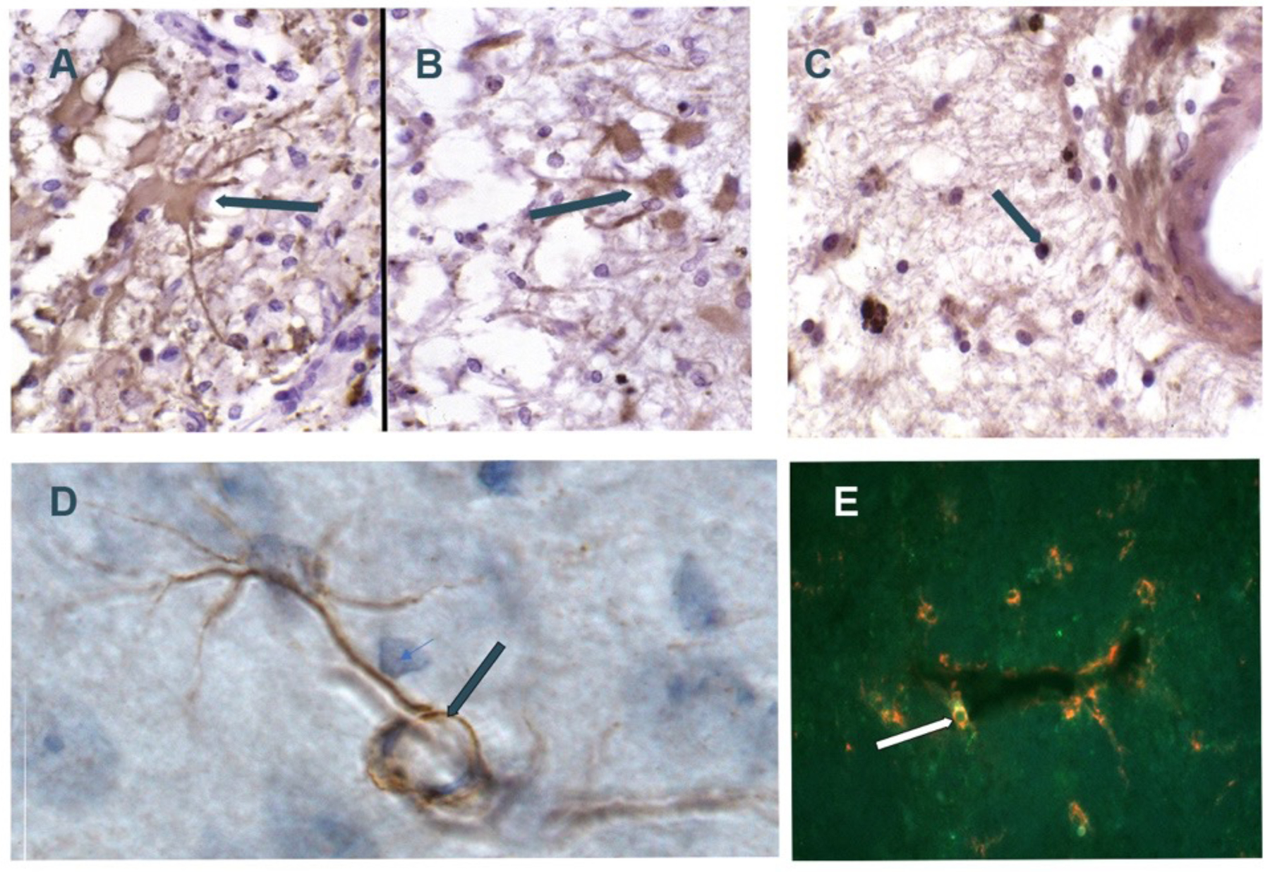
Figure 5:

Composite of MMP immunostaining. A) GFAP+ astrocyte (arrow) in patient with vascular dementia; B) Similar region as in A with astrocyte stained with MMP-2 antibody; C) PG-1+ macrophage/microglia (arrow) in patient with vascular dementia (5A,5B,5C are from Rosenberg et al46 Copyright © 2001, Wolters Kluwer Health); D) normal rat brain with MMP-2-staining astrocyte. Astrocyte foot process (arrow) wrapped around vessel; E) Rat brain dual labelled fluorescent Ox-42+ pericyte (green) and MMP-3 (red) next to vessel (arrow) (5D and 5E from Rosenberg et al70 Copyright © 2001 Elsevier Science B.V.).
