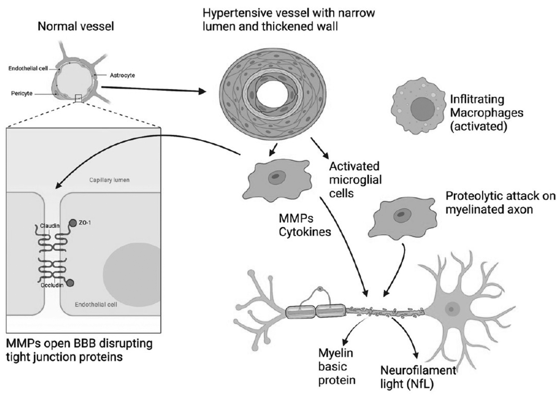
Figure 7:

Schematic illustration of by-stander demyelination mechanism in BD. A normal vessel is shown in the upper left. There are tight junction proteins between the endothelial cells that include claudin, occludin and zona occluden (zo-1). The vessel in the top center shows the effects of long-standing hypertension with narrowing of the lumen and thickening of the outer wall. Hypertensive vessels attract microglia/macrophages that attempt to remodel the vessel. These activated inflammatory cells secrete inflammatory factors, namely, MMPs and cytokines. These activated cells destroy the myelinated axons resulting in the release of myelin basic protein and neurofilament light (NfL), which can be measured in CSF and blood as a biomarker of inflammatory injury. Infiltrating macrophages (top right) can add to the proteolytic damage. (Figure created with BioRender with permission).
