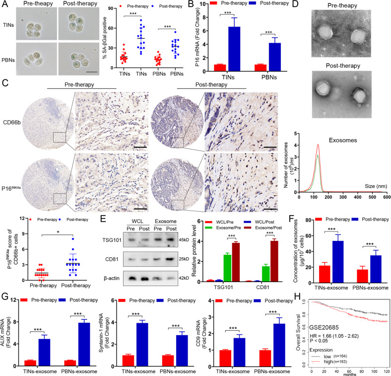
Fig. 1. Neutrophils exhibit high senescence in BC patients receiving chemotherapy with increased generation of exosomes.
A Representative image of SA-βGal staining in the TINs and PBNs extracted from BC patients (n = 15) before and after chemotherapy. Scare bars, 20 μm. B qPCR analysis of P16INK4a in the TINs and PBNs extracted from BC patients before and after chemotherapy. C Representative images of P16INK4a and CD66b staining in BC patients before and after chemotherapy (above). Scare bars, 50 μm. The P16INK4a staining score of CD66b+ cells in tumors pre- and post-chemotherapy (below). D Phenotype analysis of exosomes derived from pre-therapeutic and post-therapeutic TINs using electron microscopy and Nano Sight nanoparticle tracking analysis. E Immunoblotting of TSG101 and CD81 in whole cell lysates (WCL) and exosomes of pre-therapeutic and post-therapeutic TINs. Densitometry represents the expression of the proteins relative to β-actin. F The number of exosomes in the exosome fraction was quantified in pre-therapeutic and post-therapeutic neutrophils. G qPCR analysis of ALIX, Syntenin-1, and CD9 in the exosomes of the TINs and PBNs extracted from BC patients before and after chemotherapy. H Kaplan–Meier survival analysis showing overall survival based on the expression of senescent neutrophil gene signature in GSE20685. Data represent the mean ± SD of at least three independent experiments. *P < 0.05, **P < 0.01, ***P < 0.001.

