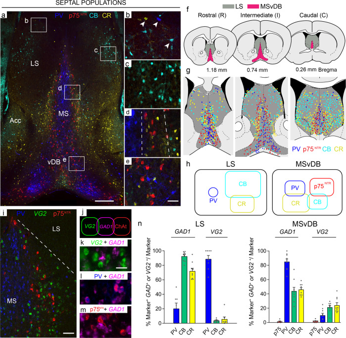
Fig. 1. Chemoarchitecture of the septal complex.
a Representative coronal sections through the adult mouse brain showing the organisation of septal nuclei. The four main neuronal subpopulations examined in this study are: PV (blue), p75NTR (red), CR (yellow) and CB (cyan). The picture was obtained by merging images from two consecutive sections. b–e Boxed areas in a are shown at higher magnification: b, c LS; d MS; e vDB. b Immunoreactivity for the three calcium-binding proteins in non-overlapping neuronal populations throughout the LS (arrowheads). c CB-expressing neurons in the LS show the typical arrangement of a nuclear structure. d The laminar organisation of the MS is shown, dashed lines indicate the borders of the layers. e p75NTR -, CB- and PV-immunoreactive neuronal populations are interspersed at the border of the MS and vDB. f Diagrams showing the three rostro-caudal levels analysed in this study. MSvDB and LS boundaries used in counts are shown (modified from ref. 71) and distribution of septal populations at rostral (R), intermediate (I) and caudal (C) levels. g Diagram showing relative abundance and overlap among the different populations of septal neurons at the three different levels examined in this study h Summary of the neuronal markers examined in this study and their overlap in the LS and the MSvDB. All overlaps indicated are <15%. i GABAergic (PV-expressing), glutamatergic and cholinergic neurons in the MSvDB. j MSvDB neurons expressing different neurotransmitters represent largely distinct populations. k–m Representative overlap between GABAergic, glutamatergic and cholinergic populations. n Quantification of co-localisation between GAD1 or VG2 cells and markers in the LS and MSvDB. n = 3 independent mice used for each marker. LS: GAD1 PV n = 7 sections; CB, CR n = 9 sections. VG2 PV, CB, CR n = 8 sections. MSvDB GAD1 p75 n = 6 sections; PV, CB, CR n = 9 sections. VG2 p75NTR n = 7 sections; PV, CB, CR n = 9 sections. Histograms show mean + SEM. Source data are provided in Supplementary Data 1. LS lateral septum, MS medial septum, Acc shell of the nucleus accumbens, vDB vertical limb of the diagonal band. Scale bars: a 200 μm, e, i 50 μm, k 10 μm.

