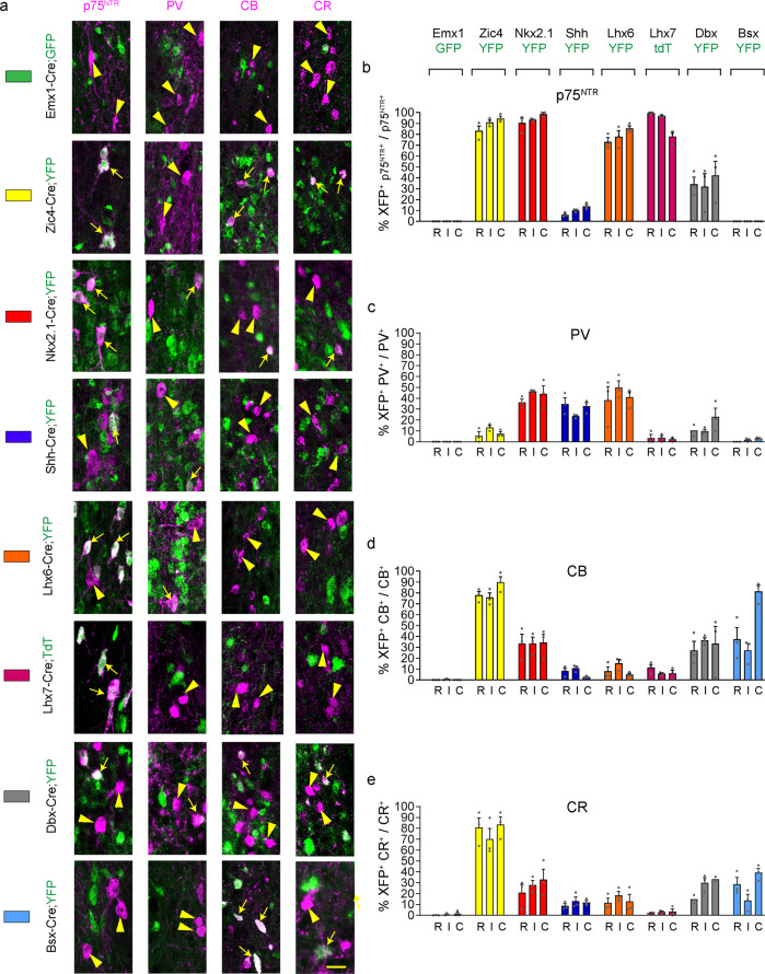
Fig. 4. Septal and extra-septal embryonic origins of medial septal neurons.
a Contribution of different forebrain domains to MS populations. Double-labelling for p75NTR, CB, CR or PV and the fluorescent reporter protein GFP/YFP/TdT in the various transgenic lines at P30. White arrows and arrowheads indicate double and single labelled cells, respectively. b–e Histograms showing the percentage of neurons double labelled for GFP/YFP/TdT over the total population. Each transgenic line is identified by a different colour as shown in a and as summarised in Fig. 2d, e. n = 3 brains per transgenic line per marker except the following where n = 2 for Dbx-Cre;YFP mice: PV R, CR R and C. Histograms show Mean + SEM. Source data are provided in Supplementary Data 1. Scale bar: 25 μm.

