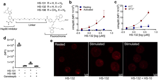
Figure 1.
T cell activation induces extracellular Hsp90 (mHsp90) expression. (a) Chemical structure of HS-131, and HS-132 active molecule, the inactive analogue HS-198 (negative control). (b) Naïve CD3+ T cells isolated from healthy human peripheral blood mononuclear cells (PBMC’s) were activated with anti-CD28 and anti-CD3 antibodies, or resting (unstimulated) for 96 h and treated with HS-132 at the indicated concentrations. Data represented as mean ± SD, 6 biological replicates. (c) Internalization of probe by CD3+ T cells at physiological (37 °C) and non-physiological (4 °C) temperatures at varying concentrations of HS-132. Data represents mean ± SD, 4 biological replicates. (d) CD3+ T cells isolated from stimulated human PBMC’s were treated with 10 µM HS-131 (active molecule) and HS-198 (inactive control) ± a tenfold excess of ganetespib. Data represents mean ± SD, 3 biological replicates. (e) Confocal analysis of HS-132 uptake in stimulated CD3+ T cells, which is blocked with treatment of the non-fluorochrome tethered Hsp90 inhibitor HS-10.

