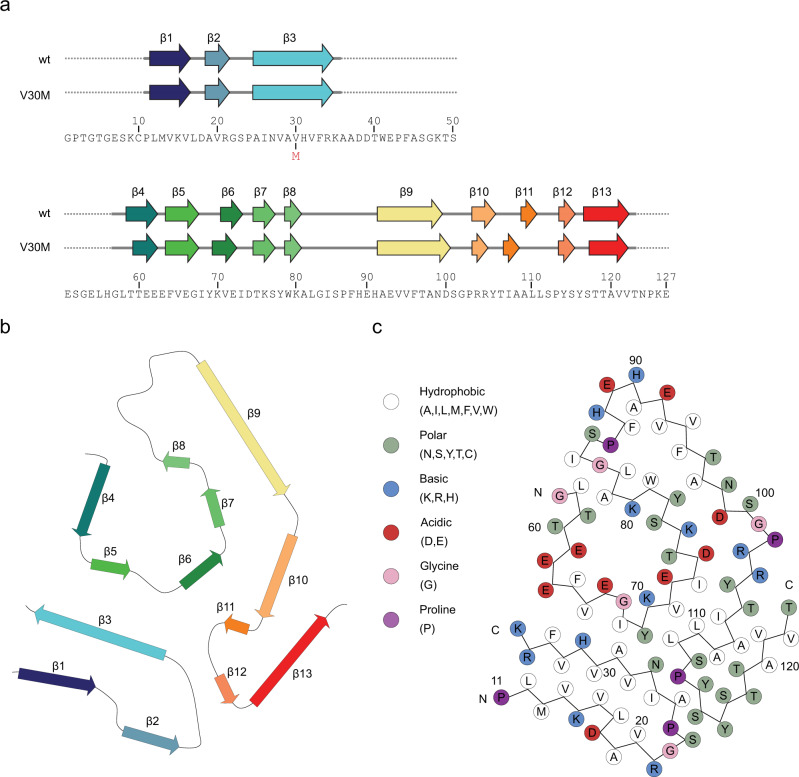
Fig. 3. β-sheet structure of the ATTR fibril.
a Amino acid sequence of the ATTRwt and the ATTRV30M variant. Above is a schematic representation of the secondary structure of the ATTRwt and the ATTRV30M variant from patient 2 23 (Protein Data bank with entry codes: 6SDZ). Arrows indicate beta-strands, continuous lines indicate resolved structure, whereas the dotted lines indicate unresolved resolved segments. The colors correspond with (b) the ribbon scheme of one protein layer inside the fibril stack. c Protein packing schematic representation of the amino acid positions.

