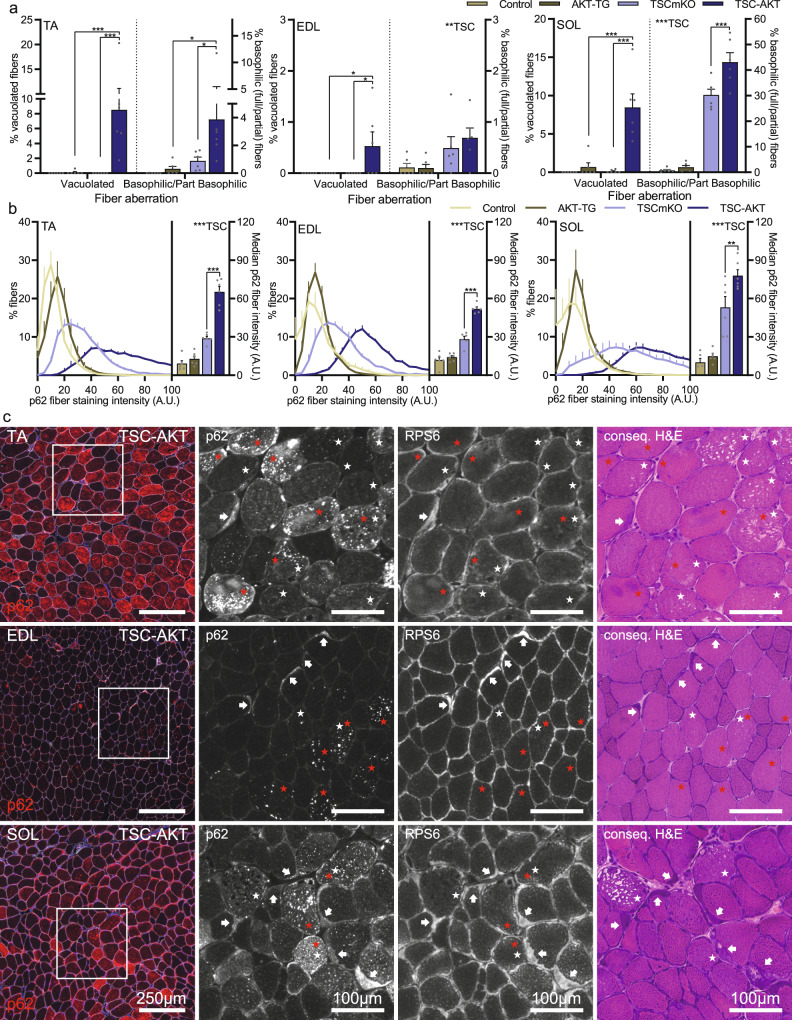
Fig. 7. Fiber vacuolation and increased p62 staining occur in parallel, but as distinct phenotypes.
a Quantification of aberrant fibers observed in Hematoxylin and eosin-stained cross sections from tibialis anterior (TA), extensor digitorum longus (EDL) and soleus (SOL) muscles collected from control, AKT-TG, TSCmKO and TSC-AKT mice. b Mean p62 fiber staining distribution (left) and median fiber staining intensity (right, column graph) for TA (left), EDL (middle) and SOL (right) muscles. c Representative images of tibialis anterior (TA), extensor digitorum longus (EDL) and soleus (SOL) cross sections showing p62 and laminin staining (left) and magnifications of p62 and RPS6 staining alongside consecutive H&E stained sections in TSC-AKT mice after 20 days of tamoxifen treatment. Vacuolated fibers (white star) and p62+ fibers (red star) frequently do not co-localize, while basophilic regions (arrows) invariably display high RPS6 staining and on occasions p62 staining. Data are presented as mean ± SEM, n = 6 per group. Two-way ANOVAs with Sidak post hoc tests were used to compare the data. *, **, and *** denote a significant difference between groups of P < 0.05, P < 0.01, and P < 0.001, respectively. For trends, where 0.05 < P < 0.10, p values are reported.

