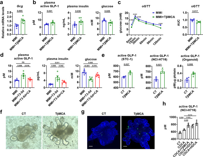
Fig. 6. Non-12α-OH FXR-antagonistic BAs mediates the T3 effect on glucose homeostasis.
a–c Relative mRNA levels of ileal Gcg (a, n = 5–6), plasma active GLP-1, plasma insulin and blood glucose levels (b, n = 5–6), oGTT (left) and the AUC for oGTT (c, n = 5) in MMI mice with or without 5 days of TβMCA treatment as indicated. d Plasma active GLP-1, plasma insulin and blood glucose levels in MMI mice treated with T3 and CA for 5 days as indicated (n = 5). e GLP-1 concentration in the supernatants of STC-1 cells (n = 3), NCI-H716 cells (n = 3) and mouse intestinal organoids (n = 4) were measured after TβMCA treatment. f Images of CT and TβMCA-treated organoids. Scale bar: 100 μm. g Representative IF staining of GLP-1 (green) and DAPI (blue) staining of mouse intestinal organoids treated with or without TβMCA. Scale bar: 70 μm. h GLP-1 concentration in the supernatants of NCI-H716 cells treated with CT, or CDCA alone or together with other FXR-antagonistic BAs as indicated (n = 3 biologically independent experiments). oGTT oral GTT. Means ± SEM are shown. P values were calculated by two-tailed unpaired Student’s t test. ns not significant. Source data are provided as a Source Data file.

