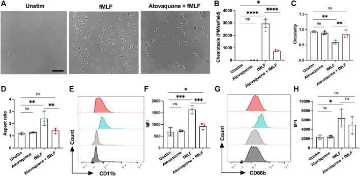
FIGURE 5.
Atovaquone negatively regulates human neutrophil chemotaxis by downregulating CD11b expression. (A–D) Freshly isolated human neutrophils were induced to migrate across permeable supports by introduction of the fMLF gradient. (A) Representative bright field images (the bar is 50 μm) and (B) Quantification of neutrophils following chemotaxis in the bottom chamber with/without pre-treatment with atovaquone. At least 7 fields per condition were quantified from 3 independent experiments that were performed in duplicates. Data are shown as an averaged value of all fields in both duplicates. (C,D) Analyses of neutrophil polarization [indexed by (C) circularity and (D) length to width ratio] following chemotaxis with/with pre-treatment with atovaquone. At least 7 fields per condition were quantified from N = 3 independent repeats in duplicates. (E–H) Human neutrophils, untreated or pre-treated with atovaquone were stimulated with fMLF (200 nM, 30 min). (E,F) Representative flow diagram and quantification of mean fluorescence intensity (MFI) of surface CD11b. (G,H) Representative flow diagram and MFI quantification of surface CD66b. N = 3 independent repeats using 3 different donors. *p < 0.05, **p < 0.01, ***p < 0.001, ****p < 0.0001, ns, not significant.

