Figure 6.
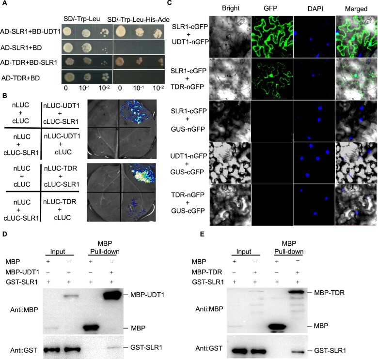
SLR1 interacts with UDT1 and TDR. A, Interaction between SLR1 and UDT1/TDR in a yeast two-hybrid assay. BD-UDT1, AD-SLR1, BD-SLR1, and AD-TDR were used as indicated. Clones that grew on SD–Trp–Leu–His–Ade medium indicate protein interaction in yeast cells. The empty AD-SLR1/BD and AD-TDR/BD were used as negative controls. B, LUC complementation imaging assay showing that SLR1 interacts with UDT1/TDR in N. benthamiana leaves. Co-infiltration of cLUC-SLR1 and nLUC-UDT1 or nLUC-TDR constructs leads to the reconstitution of the LUC signal, whereas no signal was detected when cLUC-SLR1 and nLUC, cLUC and nLUC-UDT1 or nLUC-TDR, or cLUC and nLUC were co-infiltrated. In each experiment, at least five independent N. benthamiana leaves were infiltrated and analyzed. C, BiFC assay showing that SLR1 interacts with UDT1/TDR in N. benthamiana leaves. Co-infiltration of cGFP-SLR1 and nGFP-UDT1 or nGFP-TDR leads to the reconstitution of the GFP signal, whereas no signal was detected when cGFP-SLR1 and nGFP, or cGFP and nGFP-UDT1 or nGFP-TDR were co-infiltrated. DAPI was used to visualize the nuclei. In each experiment, at least five independent N. benthamiana leaves were infiltrated and analyzed. D and E, Pull-down assay showing the interaction between UDT1/TDR and SLR1. GST-SLR1 was pulled down by MBP-UDT1 and MBP-TDR immobilized on amylose resin beads and was detected with anti-MBP and anti-GST antibody, respectively.

