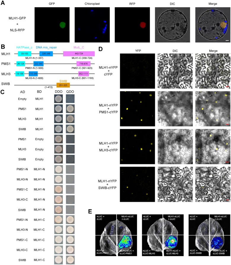
Figure 4.
Protein sub-cellular localization and protein–protein interactions. A, Sub-cellular localization of OsMLH1-GFP fusion protein in rice protoplasts. Bar = 5 μm. B, The illustration of the conserved domains of three MutL proteins and OsSWIB in rice. C, Yeast two-hybrid assay to examine interactions among MutL proteins and chromatin remodeling complex subunit SWIB. Transformed yeast cells were grown on a synthetic medium lacking Trp and Leu (DDO) or Trp, Leu, His, and Ade (QDO). AD, GAL4 activation domain; BD, GAL4 DNA-binding domain. D, BiFC analysis of the interaction between MLH1 and each of PMS1, MLH3, and SWIB. Bar = 20 μm. E, LUC complementation assay between MLH1 and each of PMS1, MLH3, and SWIB.

