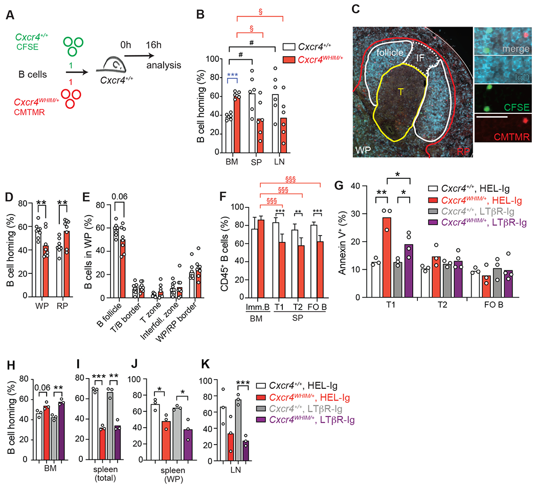
Figure 7: B cell trafficking and transitional B cell differentiation in WHIM mice.

(A) Experimental setup for adoptive transfer of fluorescently labelled Cxcr4WHIM/+ (CFSE+) and Cxcr4+/+ (CMTMR+) B cells into wildtype recipients. (B) Distribution of Cxcr4WHIM/+ and Cxcr4+/+ B cells 16 hours after adoptive transfer to C57BL6/J recipient mice. BM (BM), spleen (SP) and peripheral lymph nodes (LN). Statistical comparisons were as follows: * = for Cxcr4+/+ vs. Cxcr4WHIM/+ B cells; § = between organs of Cxcr4WHIM/+ mice. (C) Histology of splenic Cxcr4+/+and Cxcr4WHIM/+ follicles. Sections were stained for IgD (blue) and nuclei (DAPI, grey); WP = white pulp, RP = red pulp, T = T zone, IF = interfollicular zone. Scale bar is 20μm. (D) Distribution of homed Cxcr4WHIM/+ (CFSE, green) and Cxcr4+/+ (CMTMR, red) B cells in splenic red (RP) and white pulp (WP). (E) Distribution of Cxcr4+/+ and Cxcr4WHIM/+ B cells within splenic lymphoid niches. Data pooled from four experiments. For panels B, D and E, bars indicate mean, circles represent individual mice. (F) B cell chimerism 8 weeks after mixed BM transplantation of CD45.2+ Cxcr4+/+ or Cxcr4WHIM/+ and CD45.1+ cells (8:2 ration), n=5 mice/group. (G) Frequency of Annexin V+ cells in transitional (T1, T2) and mature follicular B cell subsets (FO B) in Cxcr4+/+ and Cxcr4WHIM/+ spleens from mice pre-treated with LTβR-Ig or HEL-Ig (black) for 24h. (H-K) Distribution of Cxcr4WHIM/+ and Cxcr4 B cells 24h after transfer into C57BL6/J mice pre-treated with LTβR-Ig or HEL-Ig (black) for 24h. (H) BM; (I) total spleen; (J) splenic white pulp (WP); and lymph node (K). Bars indicate mean, error bars show standard deviation. Data in all panels are representative of 2-4 independent experiments. * = p ≤ 0.05, ** = p ≤ 0.01, *** = p ≤ 0.001 by unpaired two-sided Student’s t-test.
