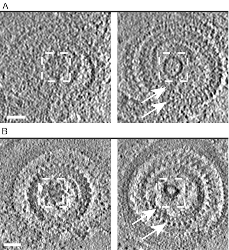
Fig. 2.

An apical vesicle is located underneath the rosette in T. gondii tachyzoites and P. falciparum merozoites. (A) Left panel is a tomographic slice showing a top view of the rosette of a Toxoplasma tachyzoite. Right panel is a different z- slice from the same tomogram showing the anterior apical vesicle (AV) and the preconoidal rings (arrows). The dashed squares mark the same area in the tomographic slice showing that the AV is positioned about 25 nm underneath the rosette. (B) As for (A) except the tomogram showing the rosette, AV, and two (out of three) apical rings of a P. falciparum merozoite. Scale bar, 50 nm.
