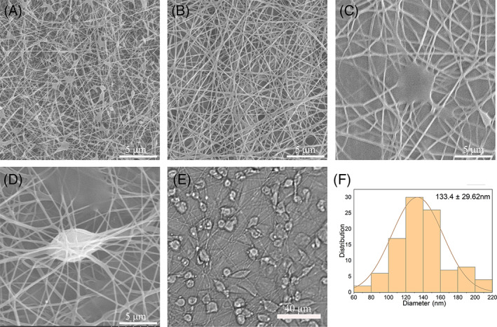
FIGURE 3.

Scanning electron microscope (SEM) images of the nanofiber membranes. (A) Fibres with 6% polyvinyl alcohol (PVA) spun with a handheld electrospinning device. (B) Fibres with 8% PVA. (C) Fibres with 10% PVA. (D) Fibres with 8% PVA and cells, spun with a handheld electrospinning device. (E) Examination of cells after cell electrospinning under a light microscope. (F) Diameter distribution of the electrospun fibres
