Fig. 2.
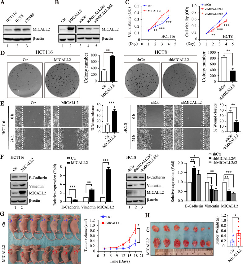
MICALL2 enhances colorectal cancer cell proliferation and migration. A The level of endogenous MICALL2 expression in HCT116, HCT8, and SW480 cell lines was detected by Western blotting assay. β-actin was used as a loading control. B The expression level of MICALL2 in stable HCT116 cell lines with MICALL2 overexpression and HCT8 cell lines with MICALL2 knockdown was detected by Western blotting assay. β-actin was used as a loading control. C MTT growth curves of stable HCT116 cell lines with MICALL2-overexpression of or HCT8 cell lines with MICALL2 knockdown. D Colony formation of stable HCT116 cell lines with MICALL2-overexpression of or HCT8 cell lines with MICALL2 knockdown. Representative photos of crystal violet–stained colonies (left) and quantifications results (right) of were shown. E Representative images and quantification of gap closure in MICALL2-overexpressed HCT116 and MICALL2-silenced HCT8 cell lines, respectively. F The expression level of E-cadherin and Vimentin in MICALL2-overexpressed HCT116 and MICALL2-silenced HCT8 cell lines detected by Western blotting assay, respectively. G Representative morphological photographs (left) of the athymic nude mice transplanted subcutaneously with HCT116 cell lines stably expressing empty vector (Ctr) or MICALL2 (MICALL2) (n = 7 per group), and dynamic volume of xenograft tumors (right) was monitored at different time points. H Photos for tumors isolated from athymic nude mice at 21 days after injection (left) and tumor weights at 21 days after injection were measured (right)

