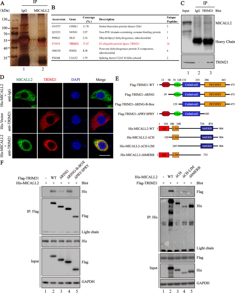
Fig. 3.
TRIM21 interacts with MICALL2. A Silver staining of proteins immunoprecipitated (IP) with MICALL2-antibody with protein extracts from CRC cells followed by identification with LC–MS/MS. The red box indicates one of the most abundant bands as compared with IgG. B List of proteins was analyzed by scaffold 4 proteome software and their information is presented. C Coimmunoprecipitations were performed to validate the interaction between endogenous MICALL2 and TRIM21 in HCT8 cells. D The colocalization of TRIM21 (red) and MICALL2 (green) in HCT8 cells was assessed by laser-scanning confocal microscopy, respectively (scale bar = 10 μm). Nuclei are stained with DAPI (blue). E Schematic representation of TRIM21, MICALL2 and its mutants. F Co-IP of His-MICALL2 with Flag-tagged TRIM21 and their truncation mutants, or Flag-TRIM21 with His- tagged MICALL2 and their truncation mutants from HEK293 cells. Cells were subjected to immunoprecipitation with α-Flag or His antibody. GAPDH is a housekeeping protein control

