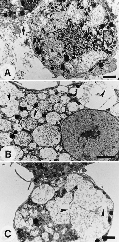
FIG. 2.
TEM studies of microglial cells cocultured with 1 mg of a lysate of A. culbertsoni per ml for 6 h. (A) A microglial cell showed numerous food vacuoles containing amoeba lysate and a lysed cell membrane (arrow). (B) The membranes of cytoplasmic vacuoles were lysed (arrowheads). (C) The lysed membranes (arrowheads) were integrated into a large one. Bars, 2.5 μm.

