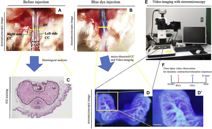
FIGURE 1.
Novel in vitro analysis of erectile responses. (A,B) After the general anesthesia and being placed in the supine position, the prepuce was removed over the penis. A measure of 0.03–0.05 ml of the PB dye solution (25 mg/ml) was slowly administered left-intracavernously through the corpus cavernosum glandis (CCG) using a 29-gauge needle. (C) Image of hematoxylin–eosin staining of the CC. Mouse proximal penis possesses two corporal units (corpus cavernosum: CC, corpus cavernosum urethra: CCU). Many sinusoidal spaces in the CC are indicated inside the dotted line. Dorsal vein (DV), dorsal artery (DA), nerve bundle (NB), sinusoidal space (SS), and urethra (U). Scale bar 200 μm. (D–F) Representative images of the PB dye-stained CC and a schematic diagram of the current experiment. The mouse with PB dye injection was harvested. The CC region was isolated by microdissections removing the prepuce, dorsal vein, artery, and nerve. Subsequently, urethra and CCU were removed to generate the CC explant. The micro-dissected CC was cultured in HBSS. The area of the cytoskeleton containing collagen fibers was efficiently marked, and the dye-stained regions were observed inside the sinusoidal space. We took time-lapse pictures every 10 s to monitor the corporal tissue responses utilizing a stereomicroscope according to the observation criteria for dynamic relaxation/contraction (see Movie.1). This study is the first direct experimental system visualizing the process of relaxation/contraction in a sinusoidal space. Time-lapse video observation criteria for dynamic relaxation/contraction. Contraction-enhancing compounds; CECs (phenylephrine and endothelin-1) and relaxation-enhancing compounds; RECs (sodium nitroprusside (SNP), acetylcholine, and prostaglandin E1). Scale bar (D) 200 μm, (D′) 100 μm. All data shown are representative results of at least three independent experiments using adult specimens from different litters (n ≥ 3).

