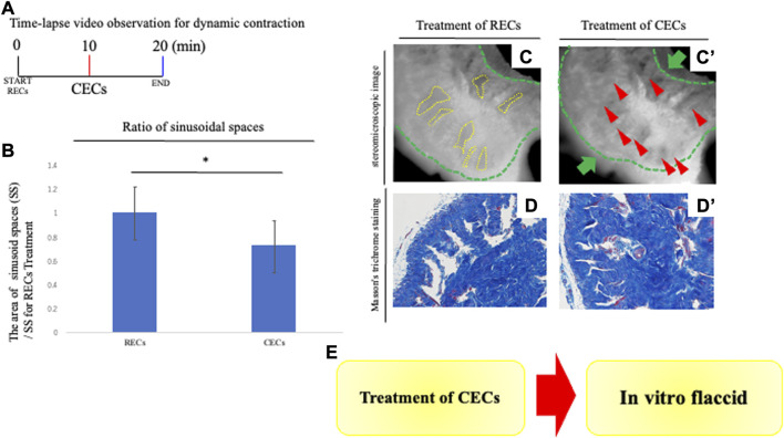
FIGURE 3.
Analysis of penile contraction responses. (A) Time-lapse video observation criteria for dynamic contraction. For analyzing the contraction of the CC, contraction-enhancing compounds (CECs) were treated to induce vasoconstriction after 10 min of the treatment of RECs. After CECs treatment, time-lapse pictures were taken for 10 min. CECs were in the final concentration of each reagent; 1.0 μM PE and 0.1 μM endothelin-1. A time-lapse video analysis was performed by ImageJ software. (B) Graph was calculated from images of the movie and indicates the relative area of sinusoidal spaces by the prominent phase of the movie. The CC with treatment of CECs was significantly decreased in its sinusoidal size compared with treatment of RECs. (C,C′) Representative 8-bit images of the PB dye-stained CC during contraction responses. Many sinusoidal spaces in the CC are indicated by yellow dotted lines. From sequential images (interval time was 10 s), the boundaries of sinusoids were marked and the sinusoidal spaces were calculated after RECs/CECs treatment by ImageJ software. We compared the area of sinusoids at each time point (the time point for RECs treatment and CECs treatment). Contraction was represented as a ratio of the treated area of sinusoid RECs to that of CECs. The whole CC (indicated by the green dotted line and arrow) was contracted, as well as the sinusoidal space. The red arrowheads showed that the collagen area contracted by the treatment of the CECs (see Supplementary Movie S2). Scale bar 100 μm. (D,D′) Image of Masson’s trichrome staining of the CC showing the collagen-rich regions. Similar to the time-lapse images, the sinusoidal spaces were reduced after treatment with CECs, and the collagen area was contracted toward the central region. Scale bar 50 μm. (E) Schema shows the reproduction of the ‘flaccid state’ by the current system. The contractive movement was in the opposite direction to that observed by the treatment of the RECs; the expanded cavernous space prominently contracted. The extended collagen regions by the REC treatment contracted in the direction of the deep arteries. The collagen regions near the deep arteries did not show significant movement when contraction was induced by the subsequent CECs. These results suggest that the “erectile state” reproduced by the REC treatment is characterized by the return to the “flaccid state.” All data shown are representative results of at least three independent experiments using adult specimens from different litters (n ≥ 3).

