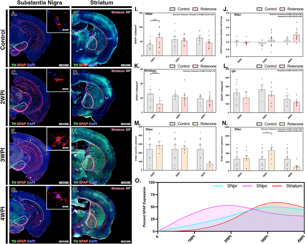
Figure 3. Rotenone exposure induces activation of astrocytes in the substantia nigra prior to the appearance of reactive A1 astrocytes in the striatum.

Activation of astrocytes (GFAP, red) within the substantia nigra (TH, green) and ST were investigated in control (A,B) and rotenone-treated mice at 2 WPI (C,D), 3 WPI (E,F) and 4 WPI (G,H). Astrocytosis/astrogliosis was quantified in the SNpc (I), SNpr (J), ST (K) and GP (L) at 2 WPI, 3 WPI, and 4 WPI. Cell number and intensity was normalized to the respective control for each region to determine overall activation patterns throughout multiple brain regions at each timepoint. The number of A1 reactive astrocytes was determined by immunolabeling and co-localization of S100β + C3 in the SNpc (M) and SNpr (N) for all timepoints. (O) Normalized GFAP intensity for the SNpr (cyan), SNpc (pink) and ST (red) was modeling for all regions at each timepoint. (N=4 mice/control group, N=7 mice/rotenone group) *p<0.05
