Figure 2:

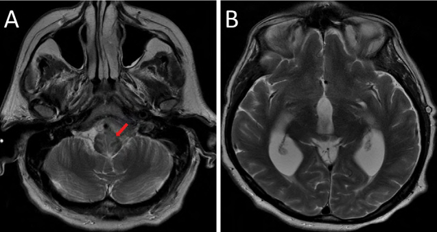
Axial T2-weighted images: (A) shows abnormal signal hyperintensity within the upper medulla, adjacent to the areas of cisternal enhancement; (B) demonstrates hydrocephalus with expansion in the caliber of the third and lateral ventricles

Axial T2-weighted images: (A) shows abnormal signal hyperintensity within the upper medulla, adjacent to the areas of cisternal enhancement; (B) demonstrates hydrocephalus with expansion in the caliber of the third and lateral ventricles