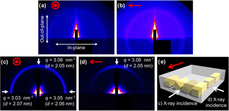
Figure 3.
2D GI-SAXS diffractograms for the polymerized A6CB film using parallel (a) or perpendicular (b) X-ray incidence to the scanning direction, respectively, and for the polymerized A0CB film using parallel (c) or perpendicular (d) X-ray incidence, respectively. (e) Possible 3D nanostructures made of smectic layers (represented by yellow layers) in the polymerized A0CB film. Red circles and arrows represent the light scanning directions.

