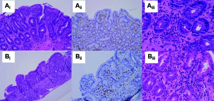
Figure 1. Duodenal histopathological features of case 1 at diagnosis and during follow-up.
(Ai) Duodenal biopsy at diagnosis showing severe villous atrophy, with normal intraepithelial lymphocyte count, as highlighted by CD3 staining (Aii) and increased eosinophilic count (Aiii). (Bi–iii) Duodenal biopsy performed 1 month after the suspension of pembrolizumab showing improvement of duodenal architecture with low-grade villous atrophy and resolution of eosinophilic mucosal infiltration.

