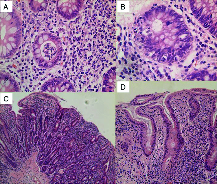
Figure 2. Colonic (A, B) and duodenal (C, D) histopathological findings in patient 2.
(A) H&E ×200. Colonic biopsy showing acute colitis with crypt abscess and mild chronic inflammation. (B) H&E ×400. Colonic biopsy showing prominent crypt cell apoptosis resembling acute graft versus host disease. (C) H&E ×100. Duodenal biopsy showing total villus atrophy with acute and chronic inflammation. (D) H&E ×200. Duodenal biopsy showing flat mucosa with cryptitis and mild chronic inflammation. Note absence of intraepithelial lymphocytes in the surface epithelium.

