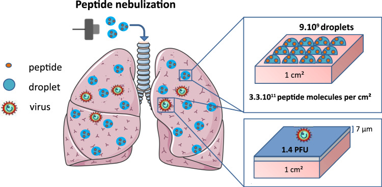
Fig. 5. Schematic presentation of the peptide and virus deposition in the lungs.
The calculation based on the estimation of the peptide dose per unit of lung internal surface, taking into consideration the number of droplets formed following the administration and the peptide molecules into the pulmonary area. Following the nebulization, 11% of the formed droplets reached the lungs (as shown in Fig. 3a), representing a density of 9×10⁹ droplets/cm2 of lung internal surface and containing 3×10¹¹ peptide molecules/cm2. Instillation of the viral inoculum (104 PFU in 5 ml) via endotracheal tube leads to the virus dispersion in the lung conductive airways in the form of 7 µm thin liquid layer (presented in blue color, covering the maximum surface of 7140 cm²). This represents 3.5 % of the total lung surface area, giving the density of infectious particles of 1.4 PFU per cm² of airways and estimated ratio between the peptide and the virus is 2 ×10¹¹ per cm2 of the lung surface.

