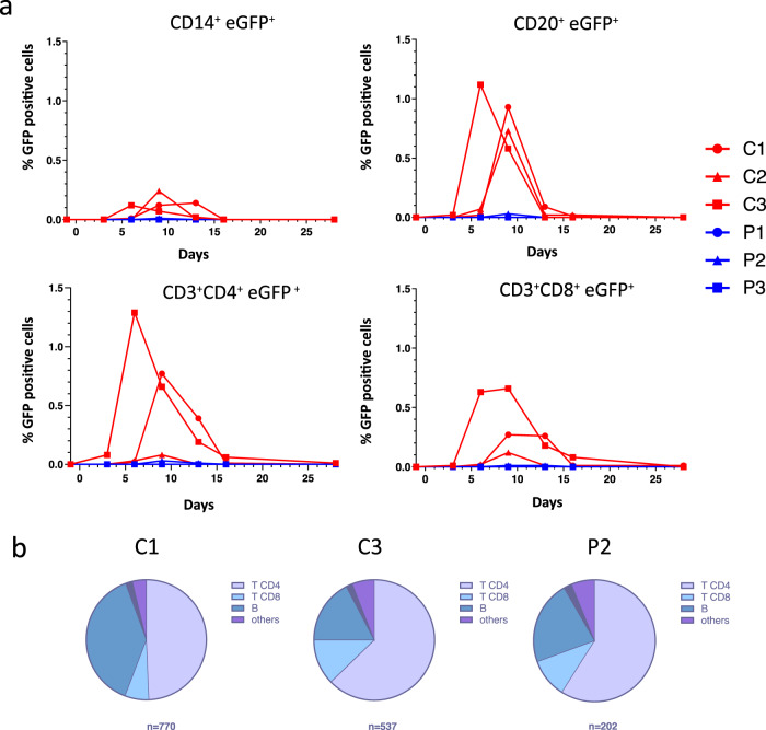
Fig. 7. Nebulization of HRC4 peptide protects PBMCs from MeV infection.
a Quantification of MeV eGFP positive cells in indicated PBMC subpopulations by flow cytometry: CD14+ monocytes, CD4+, CD8+ and CD20+ lymphocytes in MeV-infected cynomolgus monkeys by flow cytometry, following the nebulization of either 0.9% NaCl (C) or HRC4 peptide (P). CD4+ T lymphocytes were characterized as CD3+CD8-, and CD8+ T lymphocytes were characterized as CD3+/CD8+; B-lymphocytes were characterized as CD3-/CD20+ cells. b Analysis of the contribution of each lymphocyte subpopulation among infected PBMCs; results are presented as the percentage of each analyzed cell population among the infected cells on the day of peak of MeV infection (day 6 for C3 and day 9 for C1 and P2; C2 is not displayed due to a low number of infected cells). Numbers below the graphs correspond to the number of analyzed cells for each presented animal. Data were acquired on a MACSQuant® 10 flow cytometer (Miltenyi). Source data are provided as a Source Data file.

