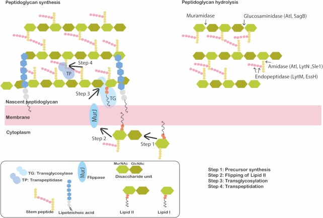
Figure 2.
Schematic representation of the S. aureus cell envelope structure. The S. aureus cell envelope is composed of a cytoplasmic membrane that is surrounded by a thick layer of peptidoglycan. For peptidoglycan synthesis (left), Lipid II units carrying disaccharides are synthesized in the cytoplasm (Step 1) and exported across the cytoplasmic membrane by a flippase (MurJ) (Step 2). Subsequently, transglycosylases (TG) insert the disaccharides into a new glycan strand (Step 3). The stem peptides of the glycan strands are then cross-linked by transpeptidases (TP) to the existing peptidoglycan matrix (Step 4). Peptidoglycan hydrolysis (right) occurs due to the action of peptidoglycan hydrolases which are classified as glycosidases (glucosaminidase and muramidase), amidases or endopeptidases. The specificity of the cleavage sites of these PGH's of S. aureus are indicated by arrows.

