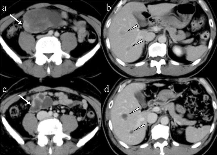
Fig. 10.
Imaging findings after imatinib therapy. A 54-year-old male with high-risk gastrointestinal stromal tumor in the small bowel was treated with imatinib. A bulky tumor with degeneration (a: arrows) and hepatic metastases (b: arrowheads) are seen in the venous phase of contrast-enhanced CT. The primary small bowel lesion decreased in size (c: arrow). Hepatic metastases show no change in size, whereas the contrast enhancement markedly decreased (d: arrowheads)

