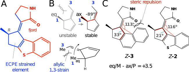
Fig. 7. Illustration of the factors contributing to absence of the stable M conformations in MTDP.
A Z-2 structure highlighting the strained unit (blue) and the clashing fjord region (red). B Pictorial illustration of the equatorial to axial relaxation imposed by the strain in a model of the ECPE unit. In the equatorial position the Me in position 1 is almost parallel/aligned with the methyl substituent in position 3. The large dihedral angle (in red) in the axial conformer is consistent with removal of the strain. C Geometrical parameters (planar and dihedral angles in red) justifying the reduced steric repulsion in Z-2 vs. Z-3. The energy difference (kcal/mol) between the M and P conformers of Z-3 is also given.

