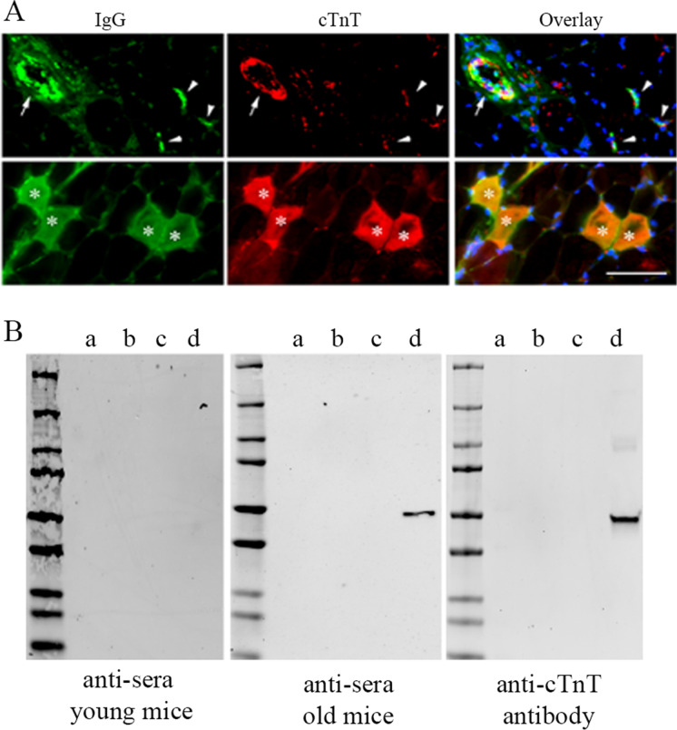
Fig. 4.
IgG anti-cTnT autoantibodies may target cTnT in the skeletal muscle of old mice. (A) Representative immunofluorescence staining image showing co-localization of IgG (stained with Alexa Fluor-488 conjugated Goat-anti-Mouse IgG) and cTnT (stained with MBS767506 and Alexa Fluor-568 conjugated Goat-anti-Rabbit) in skeletal muscle of old mice within myofibers (asterisks), at NMJs (arrowheads), and along blood vessel wall (arrow). Similar to findings in Fig. 3B and C, all most all IgG positive areas were also positive with cTnT staining. We did Goat-anti-Rabbit secondary antibody alone or with Goat-anti-Mouse secondary antibody, which did not show any signal in red (data not shown). Scale bar, 50 µm. (B) Immunoblot assay using recombinant TnT proteins detected anti-cTnT autoantibodies in serum from old mice. Pooled sera (1:100 dilution) from young or old mice (n = 3 in each age group) and anti-cTnT antibody (MBS767506, Mybiosource) were used as anti-sera or primary antibody, respectively. Recombinant cTnT was only detected by the anti-sera from old mice and anti-cTnT antibody, but not anti-sera from young mice. Recombinant proteins used for the immunoblot (20 ng per lane): a, human TnT1 protein (GTX109585-pro, GeneTex); b, mouse TnT3 isoform 8 protein with a fusion His-SUMO (Small Ubiquitin-like Modifier) tag; c, SUMO tag (GenScript); d, human cTnT isoform 11 protein (ab86685, Abcam)

