Fig. 6.
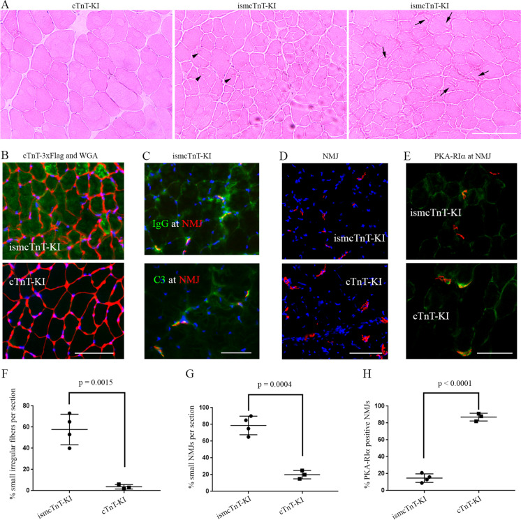
Transgenic skeletal muscle cTnT knockin mice showed signs of muscle degeneration and denervation. (A) H&E staining of tibialis anterior muscle show ismcTnT-KI mice have many smaller fibers with irregular shape, vacuoles (arrow heads), and lesions (arrows). (B) Majority of myofibers in ismcTnT-KI mice have elevated cTnT knockin overexpression (green) with irregular fiber membrane shape labeled in red with WGA (wheat germ agglutinin). (C) IgG and C3 (green) are found at NMJ (red) of ismcTnT-KI mice. (D) ismcTnT-KI mice have smaller NMJ size than the control cTnT-KI mice. (E) Compared to control cTnT-KI mice, the ismcTnT-KI mice have lower level of PKA-RIα (green) present at their NMJs (red). Images shown are representative of 4 ismcTnT-KI mice and 3 control mice analyzed. Scale bars, 50 µm. (F–H) Quantitation of (A), (D), (E), respectively. For PKA-RIα, at least 20 NMJs were counted in each mouse

