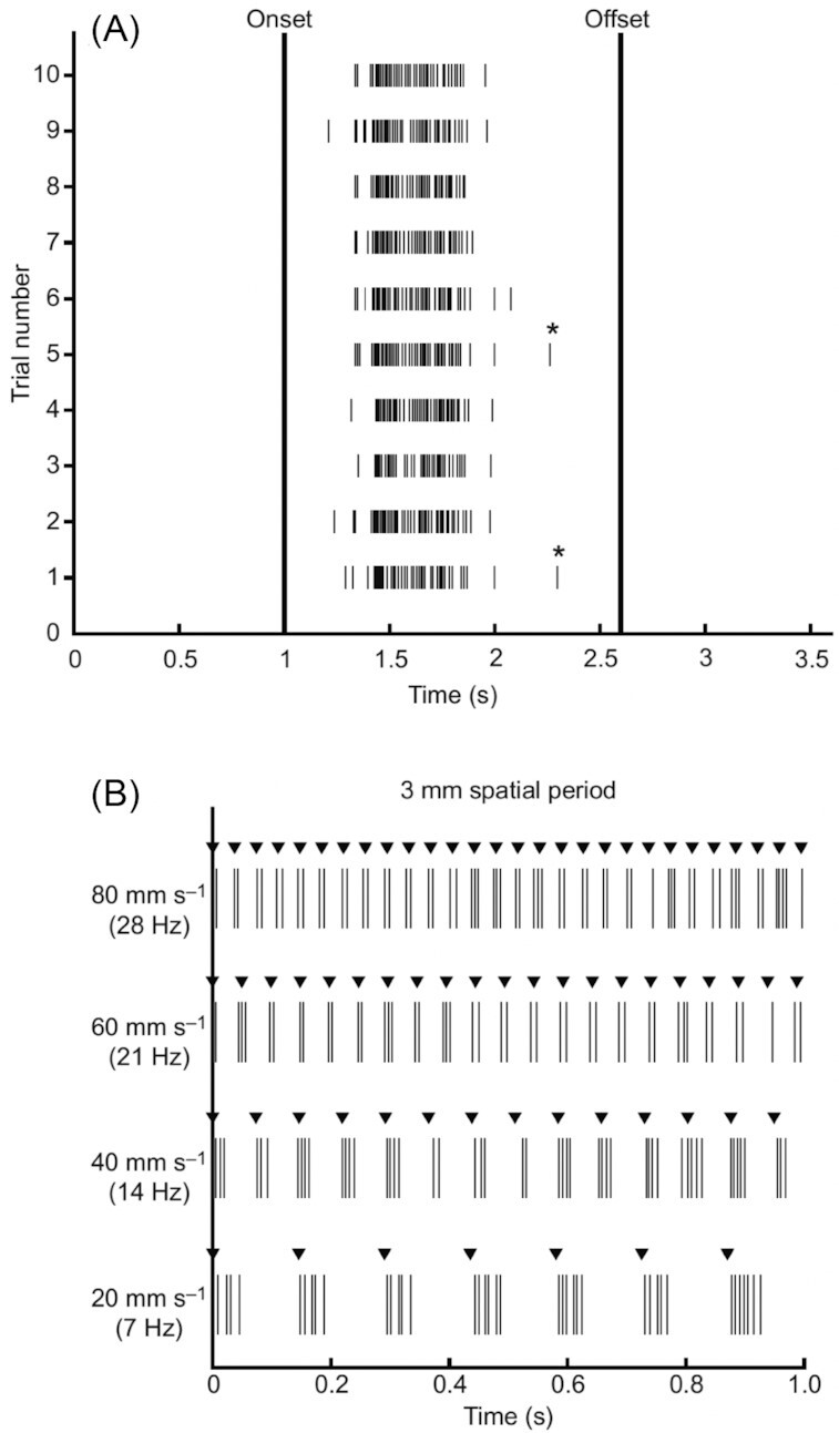
Fig. 1.

Touch sensation in the round goby, Neogobius melanostomus. Raster plots show spiking of afferent nerve fibers in response to fin ray stimulation. (A) A total of 10 trials of proximodistal brushing along an 8-mm length of a ray at 5 mms−1, passing over an afferent ending showing consistency of the response. (B) A drum with a 3-mm spatial grating rotating so that the gratings make light, brief contact with the fin, brushing proximodistally. Recordings are shown for at four speeds. The ability of the sensory afferent to sense the structure of the stimulus through this range of speeds (drum rotation rates) indicates that these fibers can faithfully report on fine substrate features. Asterisks indicate spikes excluded from analysis of spike bursts as they did not fall within the threshold for firing rate. These data show the fine spatial and temporal sensitivity and reliability of touch sensors in the pectoral fin. Reprinted from Hardy and Hale (2020)
