
Fig. 2.
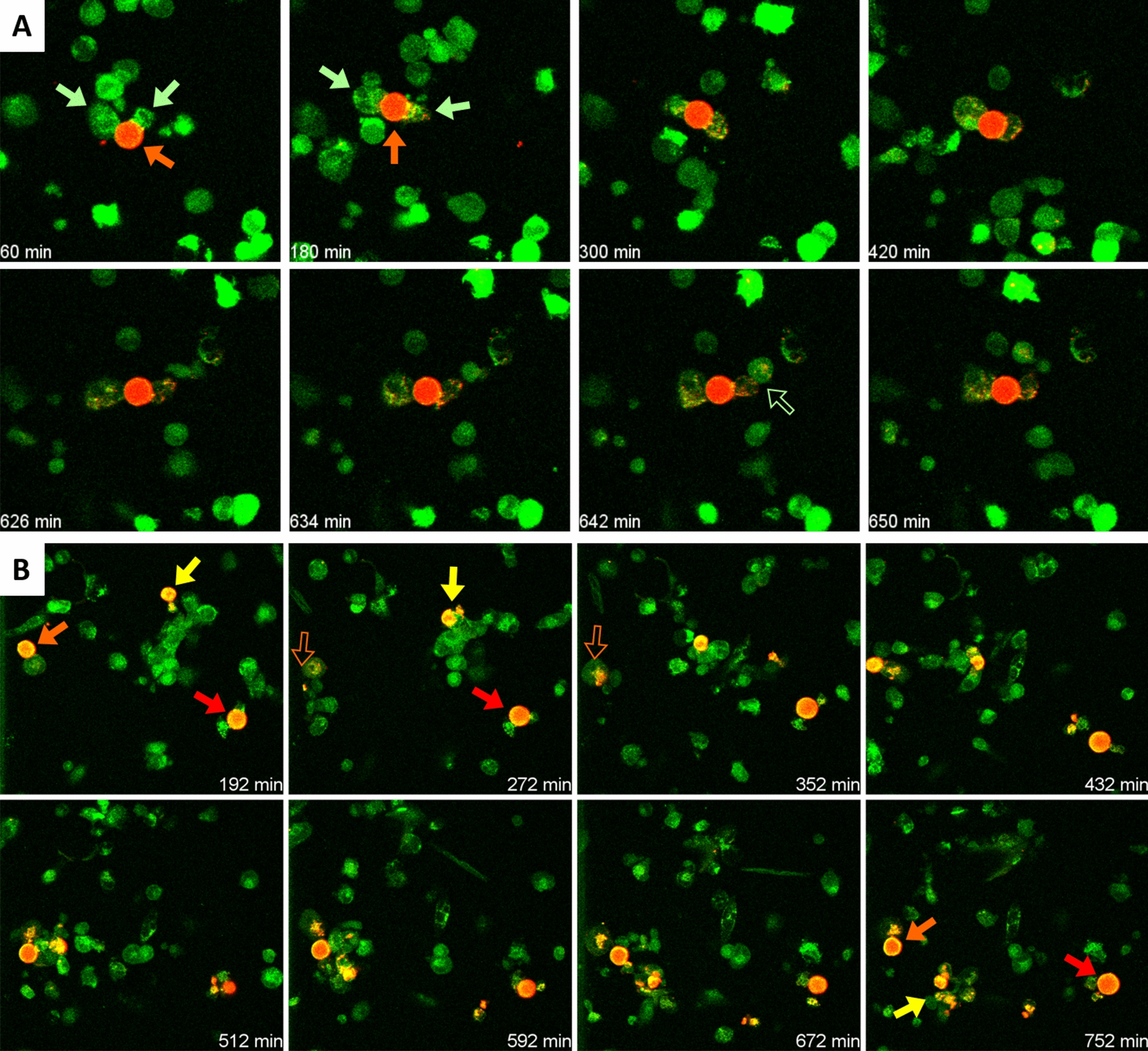
A Sequence of images from a time-lapse recording showing two THP-1 macrophages (green arrows) eroding a wasteosome opsonized with ConA (red arrow). Some spots of red fluorescence become incorporated into the macrophages. In some cases, the fluorescence is transferred from one macrophage to another one (empty green arrow). See video for details. B Sequence of images showing different macrophages interacting with three different wasteosomes (arrows). One of the wasteosomes (yellow arrow) become digested and fragmented. Empty arrows indicate that the wasteosome is out of the focus plane