Fig. 5.
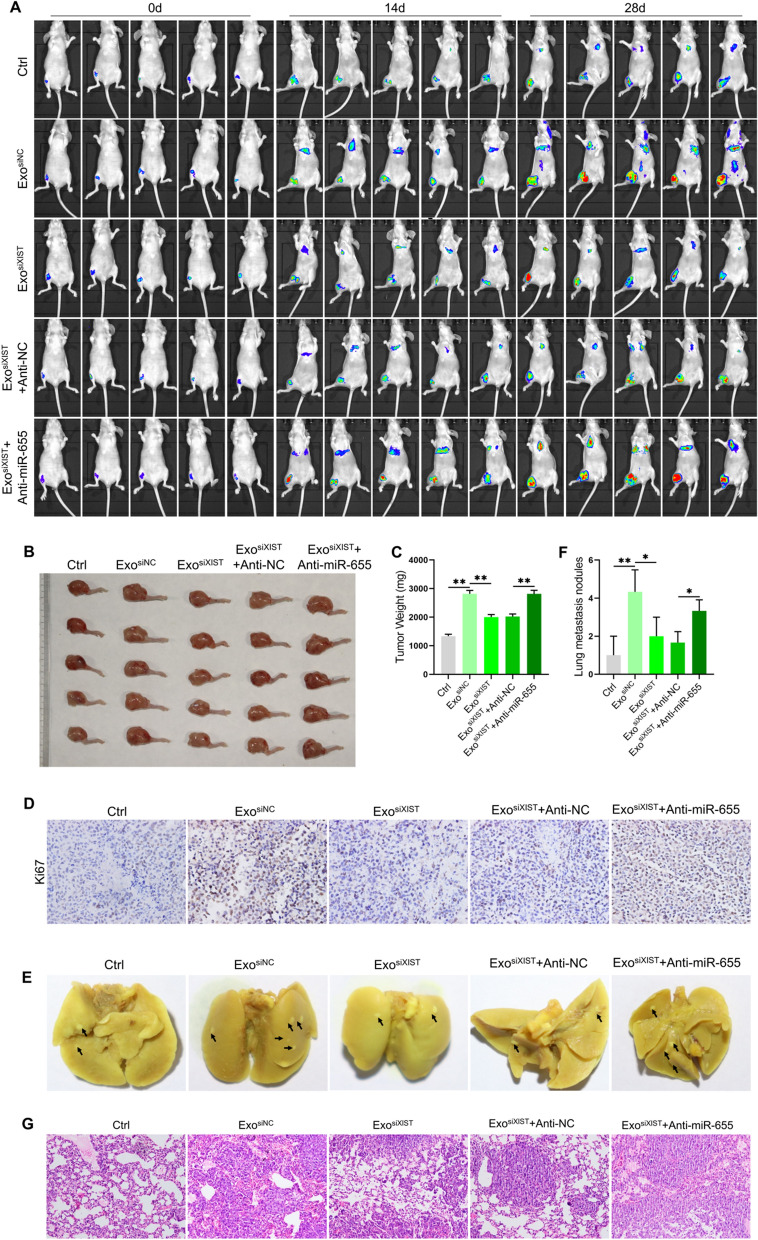
BMSCs derived exosomal XIST promoted osteosarcoma progression by binding miR-655. An in-situ osteosarcoma model was established by injecting 143B/LUC cells into the tibial bone marrow cavity of BALB/c nude mice. A The size and distribution of tumors in different treatment groups observed by in vivo imaging at 0, 14 and 28d respectively(n = 5); B The tumor size observed at 28d (n = 5); C. The weight of tumor tissues was measured (n = 5); D The positive staining of Ki67 in tumor tissue by immunohistochemical staining(n = 3); E The tumor metastasis observed after the lung tissue was dissected (n = 3); F. statistically analysis of the number of pulmonary metastases of osteosarcoma in different treatment groups(n = 3); G HE staining results to show the pathological changes of lung tissue(n = 3). *represents p < 0.05, ** represents p < 0.01

