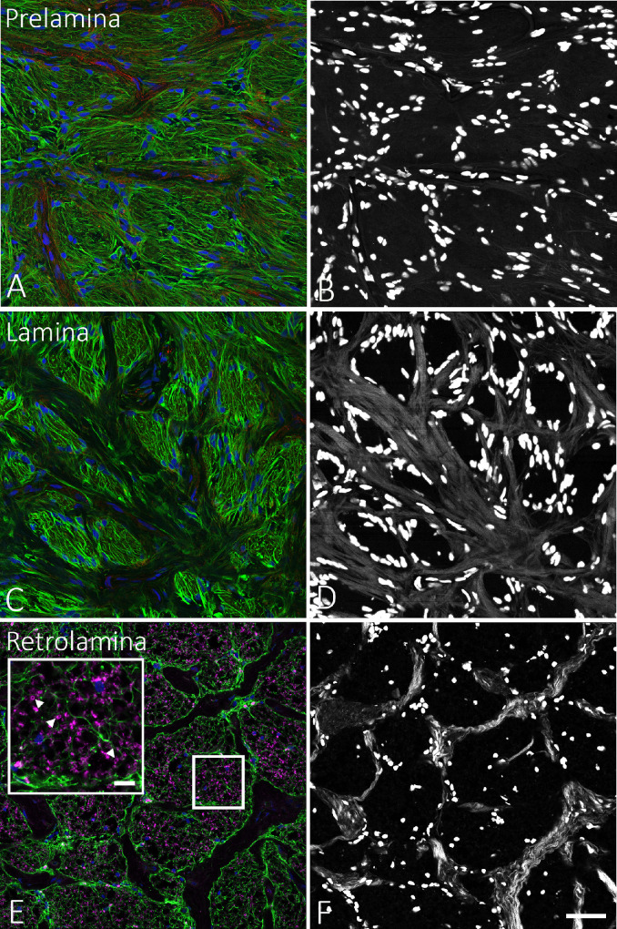
Figure 1.
Illustration of method used to identify the depth of serial sections that were cut starting in the prelamina, through the LC, to the myelinated optic nerve. (A, B) Prelamina zone, showing label for GFAP (green, A) in glial cells but without any white, collagenous beam structures in the SHG image (B). (C, D) GFAP-positive processes are seen within LC pores (C), separated in the LC by collagenous beams seen in the SHG image (D). (E) In a section that contains some portions in the retrolaminar, myelinated nerve, labeling for myelin basic protein (purple) is seen in some pores (box area is shown magnified in larger box with arrowheads pointing toward myelin basic protein label). (F) In addition, the collagenous beams in myelinated nerve have a thinner and more tortuous structure. Scale bars: 50 µm (A–F) and 5 µm (E, inset).

