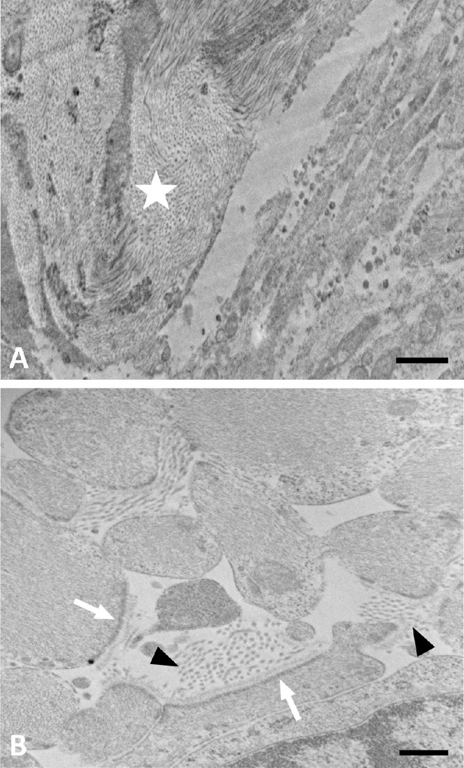
Figure 9.

TEM image of LC pore remodeling in a glaucoma. (A) Lower magnification of moderately damaged glaucoma ONH showing lamina beam (star) with collagen and elastin. Pore area at lower right shows many astrocyte processes. (B) Higher power of pore area has astrocyte processes with expanded extracellular space, containing collagen fibrils (arrowheads). Astrocytes have incomplete basement membrane with dense junctional complexes facing only those areas where basement membrane is present (white arrows). Scale bars: 1 µm (A) and 500 nm (B).
