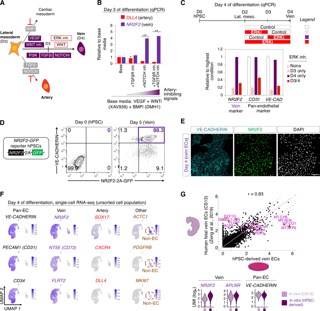
Figure 3: Efficient generation of human vein endothelial cells from hPSCs within 4 days.
A) Summary of present work
B) Dually inhibiting TGFβ and NOTCH promotes day 3 vein gene expression. Day 2 hPSC-derived lateral mesoderm was further differentiated for 24 hours with the indicated signals, including TGFβ inhibitor (SB505124, 2 μM) and/or NOTCH inhibitor (RO4929097, 1 μM); qPCR performed on day 3; *P<0.05; **P<0.01
C) VEGF/ERK activation followed by inhibition is critical for vein formation. Day 2 hPSC-derived lateral mesoderm was further differentiated for 48 hours in the presence or absence of ERK inhibitor (PD0325901, 100 nM) for the indicated durations; qPCR performed on day 4 (base media: VEGF+SB505124+RO4929097+XAV939+DMH1+AA2P for days 3–4)
D) Flow cytometry of H1 NR2F2–2A-GFP hPSCs before or after differentiation into day 5 vein ECs
E) NR2F2 and VE-CADHERIN immunostaining of H1 hPSC-derived day 4 vein ECs; scale = 100 μm
F) scRNAseq of H1 hPSCs differentiated towards vein ECs; the entire day 4 cell population was harvested without pre-selecting ECs; gene expression in loge UMI units
G) scRNAseq comparison of hPSC-derived day 4 vein ECs vs. Carnegie Stage 13 (CS13) human fetal vein ECs (Zeng et al., 2019); 6000 variable genes (top, each dot = 1 gene) or selected genes (bottom) shown

