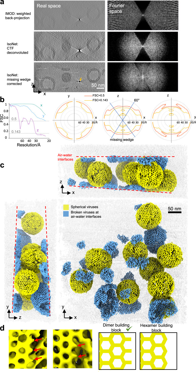
Fig. 3. IsoNet reveals lattice defects in immature HIV capsid.
a XZ slice views of the tomogram reconstructed with WBP (top), CTF deconvoluted WBP tomogram (middle), and missing-wedge corrected tomogram (bottom), with their Fourier transforms on the right. The orange arrow indicates a gold bead. b 3D FSC of the two independent isotropic reconstructions, the left panel shows the FSC along the X, Y, and Z directions. Three panels on the right show the 3D FSC at 0.5 and 0.143 cutoffs on XY, XZ, and YZ planes. c 3D rendering of the missing-wedge corrected tomogram. Dashed lines show the air-water interfaces. d Examples (left) and illustrations of the lattice edges of HIV capsids. Red arrows point out the density protrusions on the edges of hexagonal lattices.

