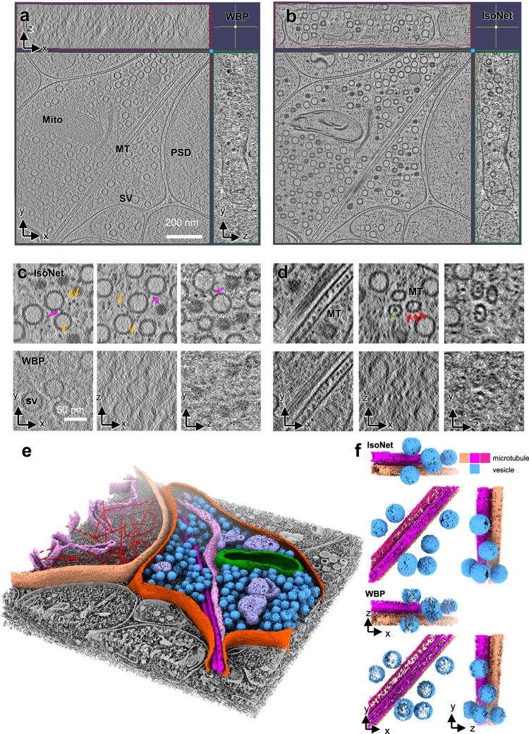
Fig. 5. IsoNet recovers missing information in the tomograms of neuronal synapses.
a, b Orthogonal slices of a synaptic tomogram reconstructed with WBP (a) and IsoNet (b). SV: synaptic vesicle; Mito mitochondria, MT microtubule, PSD postsynaptic density. c, d Zoomed-in orthogonal slices of WBP reconstruction and IsoNet-produced reconstruction. Magenta arrows: vesicle linker; Orange arrows: small cellular proteins; Green arrows: microtubule luminal particles; Red arrows: microtubule subunits. e 3D rendering of the tomogram shown in (b). f 3D rendering of a slab of tomogram with WBP reconstructions and Isotropic reconstructions, showing microtubules and vesicles.

