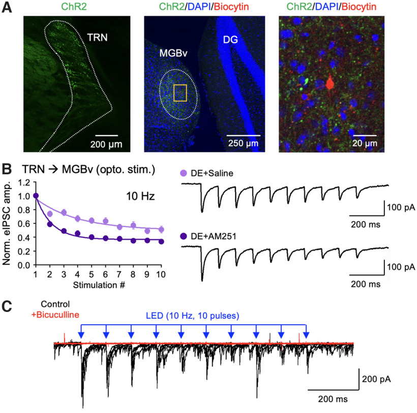
Figure 8.
CB1-R antagonist increases short-term depression of TRN inhibition onto MGBv of DE mice. A, Confocal images verifying ChR2 expression in TRN of PV-Cre mice. Left, TRN neurons expressing ChR2-YFP (green). Middle, A low-magnification tiled image showing ChR2-YFP expressing TRN axons (green) and an MGBv neuron filled with biocytin during whole-cell recording (red). The section is counterstained with DAPI (blue). DG, Dentate gyrus. Right, A zoomed-in image of the yellow boxed area shown in the middle. B, Short-term depression of ChR2-evoked IPSCs recorded from TRN to MGBv neurons in mice treated with osmotic pump containing AM251 (dark purple) or saline (light purple) during a week of DE (two-way ANOVA statistics: F(9,306) = 6.48, p < 0.0001; saline, n = 15 cells from 5 mice; AM251, n = 21 cells from 5 mice). Left, Comparison of the normalized eIPSC amplitude recorded during a 10 Hz train (10 pulses) optical stimulation. Plot, Mean ± SEM. Right, Example eIPSC traces from saline control and AM251-treated groups. C, ChR2-eIPSCs are blocked by bath application of bicuculline (10 μm). Overlay of 5 consecutive traces before (black) and after (red) bicuculline application. Bicuculline blocked both evoked and spontaneous IPSCs.

