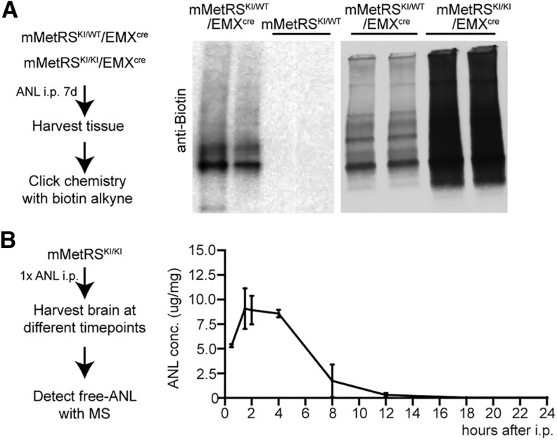
Figure 4.
Optimization of ANL protein labeling in vivo. A, Comparison of ANL incorporation in heterozygote or homozygote mMetRSKI/WT/EMX and mMetRSKI/KI/EMX mice and mMetRS/WT littermates. Left, Schematic of the protocol. Animals were treated with a daily dose of ANL (830 mg/kg, i.p.) for 1 week, and the brain was harvested 18–20 h after the last ANL treatment. Tissue was processed for click chemistry with biotin-alkyne. Middle, Western blots comparing ANL-biotin-labeled proteins from mMetRSKI/WT/EMXcre and mMetRSWT/WT littermates. No ANL incorporation into proteins is observed in the absence of cre-induced mMetRS expression. Right, Western blots showing more ANL-biotin label in comparable amounts of protein samples from mMetRSKI/KIEMXcre mice compared with mMetRSKI/WT/EMXcre mice. B, Analysis of brain ANL levels after intraperitoneal injection by mass spectrometry. Left, Schematic of the experiment. mMetRS mice received 1 dose of ANL intraperitoneally and brains were harvested at different timepoints after injection. Free ANL in the brain was detected by MS. Right, ANL levels in the brain over time [mean ± SEM; N = 4 mice (2 males and 2 females) paired t test with Welsh's correction]. ANL concentration was calculated by the amount of ANL in ug normalized to the amount of brain tissue analyzed in mg.

