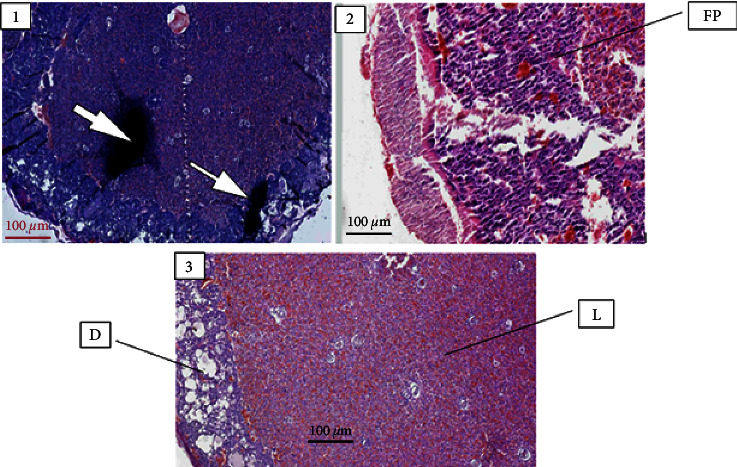
Figure 3.

Photo micrograph depicting the following: (1) a calcified placental tissue (arrow) from rats treated with 1000 mg/kg of Embelia schimperi, (2) fibroprulent tissue (FP) from rats treated with 500 mg/kg, and (3) normal histology of placenta from the control rats; decidual layer (D), labyrinthine zone (L).
