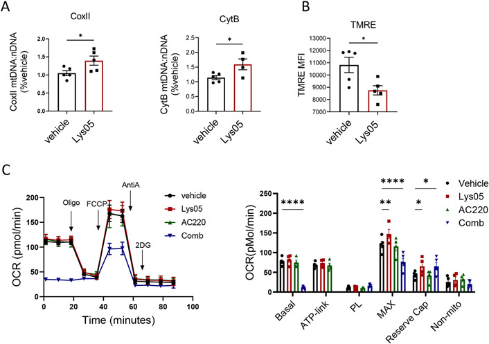
Figure 7. Reduced mitochondrial respiration in AML stem cells with tyrosine kinase inhibition in combination with autophagy inhibition.
A: Mitochondrial DNA (mtDNA) from Lys05 and vehicle treated LSK cells(n=5) was quantified based on ratio of mt-CoxII or mt-CytB to nuclear DNA. B: Mitochondrial membrane potential was measured by TMRE labeling in LSK cells from Lys05 and vehicle treated mice(n=5). C: Extracellular flux analysis for oxygen consumption rate (OCR) was performed on c-Kit enriched cells obtained from FLT3-ITDki /Mx1-Cre Tet2f/f leukemic mice after 2 weeeks of indicated drug treatment (n=4-5). A representative plot and a graph of compiled data are shown. Significance values: *p<0.05; **p<0.01; ***p<0.001; ****p<0.0001; ns, not significant. Results represent mean ± SEM of multiple replicates.

