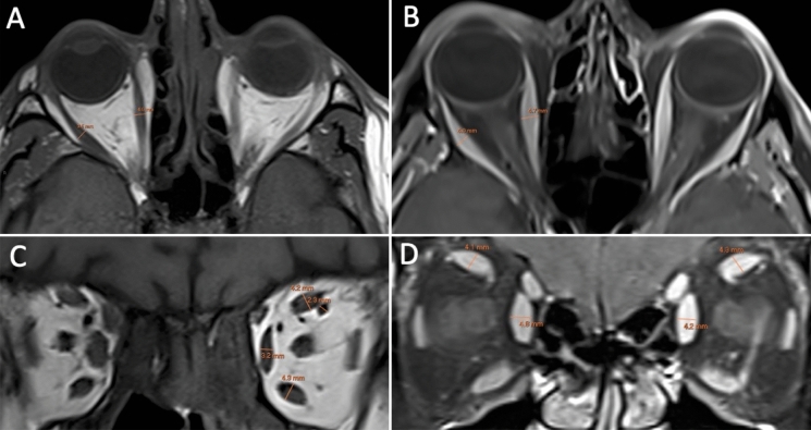
Fig. 1.
Axial T1-weighted MRI (A) and fat-suppressed contrast enhanced T1-weighted MRI (B) showing the measurements of the right medial and lateral rectus muscles perpendicular to the muscle belly. Coronal T1-weighted MRI (C) showing the measurements of the superior muscle group, medial rectus, inferior rectus and superior ophthalmic vein. Fat-suppressed contrast enhanced T1-weighted MRI (D) showing the measurements of the medial rectus and superior muscle group

