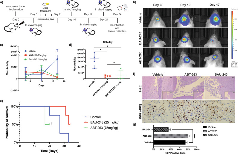
Fig. 7. BAU-243 significantly inhibited in vivo tumor growth.
a Summary of experimental design. 1 × 10⁵ Firefly Luciferase expressing U87MG GBM cells inoculated into the brains of NOD/SCID Gamma mice with a stereotactic frame. From day 3 through day 7, each treatment group (n = 4) received 5 doses of either vehicle, ABT-263 (75 mg/kg), or BAU-243 (25 mg/kg). Tumor growth was visualized and followed by in-vivo imaging, and on day 34 experiment was ended to obtain brain tissues. b In vivo imaging of Vehicle, ABT-263 (75 mg/kg), and BAU-243 (25 mg/kg) treated mice. Representative one mouse from each group is shown. c Tumor growth Fluc activity of Vehicle, ABT-263 (75 mg/kg), and BAU-243 (25 mg/kg) treated mice. d Fluc activity of each animal in each group on the 17th day. e Kaplan Meier curve of the probability of survival for each group. † Remaining two animals from ABT-263 treated group had to be euthanized due to ethical concerns. f H&E and Ki67 staining of brain sections from each treatment group. H&E images were shown as both 4X and 10X magnifications. Ki67 images were shown at 40X magnification. g Percentage of Ki67 positive cells. The percentile of Ki67 positive cells relative to all stained cells is calculated from three independent images for each group. Data are expressed as mean ± SD. Statistical analysis was performed using ordinary one-way ANOVA, together with Dunnett’s multiple comparisons test. *p < 0.05, ****p < 0.0001.

