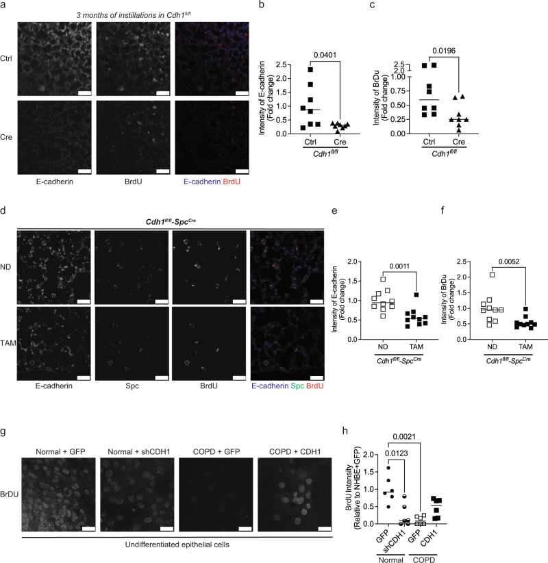
Fig. 3. Knockdown of E-cadherin decreases the regeneration of epithelium in the mice lungs.
Regeneration of epithelium was assessed by BrdU staining. (a) Representative image at 10X (scale bar of 50 µm) of Cdh1fl/fl mice instilled with adeno-Cre recombinase (Ad5CMVCre-eGFP) to knockdown E-cadherin for 3 months shows decreases in BrdU as compared to Cdh1fl/fl instilled with adeno-Ctrl (Ad-5CMVeGFP). Decreases in the fluorescence intensity of b E-cadherin and c BrdU in Cdh1fl/fl mice instilled with adeno-Cre as compared to Cdh1fl/fl mice instilled with adeno-Ctrl. Data is generated from eight mice. To knock down E-cadherin in the AT2 cells of mice lungs, Cdh1fl/fl-SpcCre mice were fed tamoxifen (TAM) for 30 days. These were compared to Cdh1fl/fl-SpcCre mice receiving a normal chow diet (ND). In vivo knockdown of E-cadherin in Cdh1fl/fl-SpcCre mice show decreases in BrdU expression as observed in the d representative images at 10X (scale bar of 25 µm), with decreases in the intensity of e E-cadherin and f BrdU in AT2 cells. Data is generated from five mice. Undifferentiated normal basal epithelial cells were transfected with Ad-GFP-U6-h-CDH1-shRNA to knock down E-cadherin, and undifferentiated COPD cells were transfected with Ad-GFP-U6-h-CDH1 to overexpress E-cadherin. We compared to respective undifferentiated Normal/COPD with Ad-GFP. g Representative image at 40× (scale bar of 25 µm) and h quantification showing decreased BrdU intensity in the normal epithelium with E-cadherin knockdown (Normal + shCDH1) and COPD at baseline (COPD + GFP) as compared to Normal + GFP. Also, COPD with overexpressed E-cadherin (COPD + CDH1) demonstrates increased BrdU intensity compared to COPD + GFP. Data are expressed as median bars and generated from three inserts from two donors.

