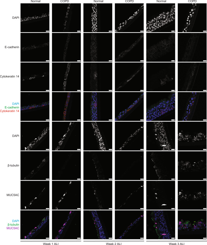
Fig. 4. Knockdown of E-cadherin decreases epithelial regeneration in human epithelial cells.
Immunofluorescence at 40× (scale bar of 50 µm) of COPD human bronchial epithelial cells differentiated at week one to three of air–liquid interface (ALI) show decreased expression of E-cadherin and β-tubulin (ciliated cells marker) expression, increase expression of MUC5AC (goblet cell marker), without any changes in Cytokeratin 14 (Basal cell marker), as compared to non-diseased human bronchial epithelial (normal) cells.

