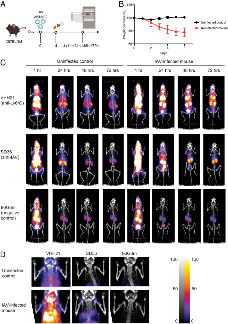
Fig. 3.
Ly6C/G-positive cells accumulate in the lung, and their presence correlates with weight loss and virus burden in the lung. (A) Schematic showing the experimental outline. (B) Graphs showing the percent weight-loss progression after influenza A virus (IAV) infection. (C) Representative (n = 3) immune-PET images of IAV-infected mice injected with 89Zr-VHH21, 89Zr-SD36, and 89Zr-96G3m at the indicated time points. (D) Images focused on the upper thoracic region (lungs) from (C) at the 48-h time point. Scale at Right indicates least intense signal to most intense.

