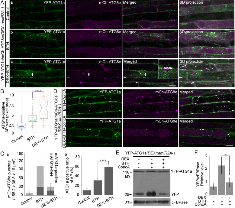
Fig. 6.
ATG1a recruited and accumulated on the enlarged autophagosomal structures in ORP2A KD plants. (A) Seven-day-old transgenic YFP-ATG1a/mCh-ATG8e/DEX::amiR2A-1 seedlings were treated with/without BTH for 6 h prior to confocal imaging and 3D projection (a and b). ΔZ = 0.8 μm, 20 stacks. For DEX+BTH treatment (c), DEX was pretreated for 48 h before BTH treatment, followed by confocal imaging and 3D projection. Arrows indicate examples of YFP-ATG1a–positive autophagic punctae containing mCh-ATG8e and enlargement in the box. (Scale bar, 10 μm.) (B) Quantification of the sizes of mCh-ATG8e–positive autophagosomes colocalized with YFP-ATG1a in A. Fifty punctae were measured using ImageJ from at least 20 root cells in each of the treatments shown in A. Error bars indicate ±SD; ****P ≤ 0.0001, Student’s t test. (C) Quantification of mCh-ATG8e punctae (a) and colocalization rate of ATG1a-positive mCh-ATG8e punctae (b) in A. (D) Seven-day-old transgenic YFP-ATG1a/mCh-ATG8e/DEX::amiR2A-1 seedlings were treated with BTH and ConcA with/without DEX pretreatment as indicated, followed by confocal imaging. (Scale bar, 10 μm.) (E) Immunoblot analysis of YFP-ATG1a protein turnover in YFP-ATG1a/DEX::amiR2A-1 seedlings. Seven-day-old transgenic YFP-ATG1a/DEX::amiR2A-1 seedlings were treated with/without BTH and ConcA for 6 h or pretreated with DEX for 48 h as indicated, followed by protein extraction and immunoblot detection with various antibodies. YFP-ATG1a and free YFP protein levels were detected by GFP antibodies. cFBPase antibodies were used as protein loading control. (F) Quantification of free YFP level normalized to cFBPase in E. Error bars indicate ±SD; *P ≤ 0.05, Student’s t test.

